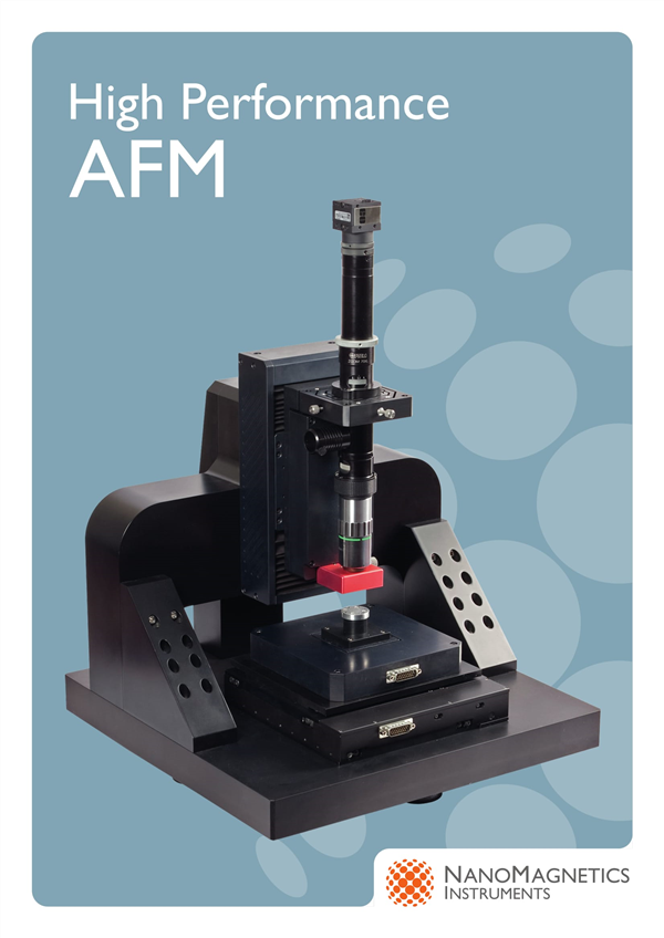
Concept: Atomic Force Microscope (AFM), an analytical instrument that can be used to study the surface structure of solid materials, including insulators. It studies the surface structure and properties of a substance by detecting the extremely weak interatomic interaction between the surface of the sample to be tested and a micro force-sensitive element. A pair of weakly sensitive micro cantilevers are fixed at one end, and the tiny tip of the other end is close to the sample, at which point it will interact with it, and the force will cause the microcantilever to deform or move. When scanning the sample, the sensor is used to detect these changes, and the force distribution information can be obtained, thereby obtaining the surface topography structure information and the surface roughness information at a nanometer resolution. Principle: The atomic force microscope (AFM) uses the microcantilever to sense and amplify the force between the sharp probe on the cantilever and the atom of the sample to be tested, and achieves the atomic resolution. Because the atomic force microscope can observe both the conductor and the non-conductor, it can make up for the shortcomings of the scanning tunneling microscope. Atomic force microscopy was invented by Gerd Binning of the IBM Research Center in Zurich in 1985. The purpose is to make non-conductors similar to scanning probe microscopy (SPM). The biggest difference between atomic force microscopy (AFM) and scanning tunneling microscopy (STM) is that it does not use electron tunneling effects, but detects the contact between atoms, atomic bonding, van der Waals force or Casimir effect. The surface characteristics of the sample. Composition: It consists mainly of a microcantilever with a tip, a microcantilever motion detection device, a feedback loop that monitors its motion, a piezoelectric ceramic scanning device that scans the sample, and a computer-controlled image acquisition, display, and processing system. The microcantilever motion can be detected by optical methods such as tunnel current detection or optical methods such as beam deflection method and interference method. When the tip and the sample are close to each other and there is a short-range mutual repulsive force, the surface repulsive force can be detected to obtain a surface atomic-level resolution image. The resolution is also at the nanoscale level. AFM measurement has no special requirements for samples, and can measure solid surface, adsorption system and so on. Advantages: Atomic force microscopy has many advantages over scanning electron microscopy. Unlike electron microscopy, which only provides two-dimensional images, AFM provides a true three-dimensional surface map. At the same time, AFM does not require any special treatment of the sample, such as copper or carbon, which can cause irreversible damage to the sample. Third, the electron microscope needs to be operated under high vacuum conditions, and the atomic force microscope can work well under normal pressure even in a liquid environment. This can be used to study biological macromolecules, even living biological tissues. Atomic force microscopy has a wider applicability because it can observe non-conductive samples compared to Scanning Tunneling Microscope. Scanning force microscopes, which are currently widely used in scientific research and industry, are based on atomic force microscopy. Disadvantages: Compared with scanning electron microscopy (SEM), AFM has the disadvantage that the imaging range is too small, the speed is slow, and it is too much affected by the probe. Structure: In the Atomic Force Microscopy (AFM) system, it can be divided into three parts: the force detection part, the position detection part, and the feedback system. Force detection part: In an atomic force microscope (AFM) system, the force to be detected is the van der Waals force between atoms and atoms. Therefore, in this system, a tiny cantilever is used to detect the amount of change in force between atoms. The microcantilever is typically made of a silicon or silicon nitride wafer that is typically 100 to 500 μm long and approximately 500 nm to 5 μm thick. The tip of the microcantilever has a sharp tip that detects the interaction between the sample and the tip. The tiny cantilever has certain specifications, such as length, width, modulus of elasticity, and the shape of the tip. These specifications are chosen according to the characteristics of the sample and the mode of operation, and different types of probes are selected. Position detection part: In the atomic force microscope (AFM) system, when the needle tip interacts with the sample, the cantilever cantilever swing. When the laser is irradiated on the end of the microcantilever, the position of the reflected light is also The cantilever swings and changes, which causes the offset to occur. In the entire system, the laser spot position detector is used to record and convert the offset into an electrical signal for signal processing by the SPM controller. Feedback system In an atomic force microscope (AFM) system, after the signal is taken in via a laser detector, this signal is used as a feedback signal in the feedback system as an internal adjustment signal and drives a scan usually made of piezoceramic tubes. The device is moved appropriately to maintain a certain force between the sample and the tip. to sum up The AFM system uses a scanner made of piezoceramic tubes to precisely control small scan movements. Piezoelectric ceramics are a peculiar material. When a voltage is applied to the symmetrical two end faces of a piezoelectric ceramic, the piezoelectric ceramics are elongated or shortened in a specific direction. The size of the elongation or shortening is linear with the magnitude of the applied voltage. That is, the micro telescopic of the piezoelectric ceramic can be controlled by changing the voltage. Usually three piezoelectric ceramic blocks representing the X, Y, and Z directions are formed into a tripod shape, and the X-Y direction is controlled to achieve the purpose of driving the probe to scan on the sample surface; by controlling the Z-direction piezoelectric ceramic expansion and contraction The purpose of controlling the distance between the probe and the sample is achieved. The atomic force microscope (AFM) combines the above three parts to present the surface characteristics of the sample: in an atomic force microscope (AFM) system, a tiny cantilever is used to sense the interaction between the tip and the sample. This force causes the micro cantilever to oscillate, and then the laser is used to illuminate the end of the cantilever. When the swing is formed, the position of the reflected light is changed to cause an offset, and the laser detector records the offset. The signal at this time will also be sent to the feedback system to facilitate proper adjustment of the system, and finally the surface characteristics of the sample will be presented as images. Mode of operation: The mode of operation of an atomic force microscope is categorized in the form of a force between the tip and the sample. There are three main modes of operation: contact mode, non-contact mode, and tapping mode. Contact mode Conceptually, contact mode is the most direct imaging mode of AFM. AFM During the entire scanning imaging process, the probe tip is always in close contact with the sample surface, and the interaction force is the repulsive force. When scanning, the force exerted by the cantilever on the tip of the needle may damage the surface structure of the sample, so the force ranges from 10 - 10 to 10 - 6 N. If the surface of the sample is tender and cannot withstand such forces, it is not advisable to use the contact mode to image the surface of the sample. Non-contact mode When the non-contact mode detects the surface of the sample, the cantilever oscillates at a distance of 5 to 10 nm above the surface of the sample. At this time, the interaction between the sample and the tip is controlled by Van der Waals force, usually 10 - 12 N, the sample will not be destroyed, and the tip will not be contaminated, which is especially suitable for studying the surface of soft objects. The disadvantage of this mode of operation is that it is very difficult to achieve this mode in a room temperature atmosphere. Because the surface of the sample inevitably accumulates a thin layer of water, it creates a small capillary bridge between the sample and the tip of the needle, drawing the tip of the needle together with the surface, thereby increasing the pressure on the surface of the tip. Tap mode The tapping mode is between the contact mode and the non-contact mode and is a hybrid concept. The cantilever oscillates above its surface at its resonant frequency, and the tip merely periodically contacts/knocks the surface of the sample periodically. This means that the lateral forces generated when the tip touches the sample are significantly reduced. Therefore, the AFM tapping mode is one of the best choices when testing soft samples. Once the AFM begins to scan the sample, the device is then used to enter the system, such as surface roughness, average height, maximum distance between peaks and valleys, for surface analysis. At the same time, the AFM can also perform force measurement and measure the degree of bending of the cantilever to determine the force between the tip and the sample. Comparison of three modes Contact Mode: Advantages: The scanning speed is the only hard sample with a significant change in the vertical direction of the AFM that can obtain the "atomic resolution" image, and is sometimes more suitable for scanning with the Contact Mode. Disadvantages: Lateral forces affect image quality. In the air, the adhesion between the tip and the sample is large because of the capillary action of the adsorbed liquid layer on the surface of the sample. The combined forces of lateral force and adhesion result in reduced spatial resolution of the image, and scratching the sample with the tip can damage soft samples (eg, biological samples, polymers, etc.). Non-contact mode: Advantage: No force acts on the surface of the sample. Disadvantages: Due to the separation of the tip from the sample, the lateral resolution is low; in order to avoid contact with the adsorption layer, the tip of the needle is glued, and the scanning speed is lower than the Tapping Mode and the Contact Mode AFM. It is usually only used for samples that are very afraid of water. The layer of adsorbed liquid must be thin. If it is too thick, the tip of the needle will fall into the liquid layer, causing the feedback to be unstable and scraping the sample. Due to the above shortcomings, the use of on-contact mode is limited. Tap mode: Advantages: The effect of lateral forces is well eliminated. The force caused by the adsorbed liquid layer is reduced, the image resolution is high, and it is suitable for observing soft, brittle, or adhesive samples without damaging the surface. Disadvantages: Slower than Scan Mode AFM. Mobile Flipchart U Stand,Magnetic Whiteboard Sheets For Board,Magnetic Glass Flipchart Easel Board,Magnetic Flipchart Easel Board Dongguan Aoxing Audio Visual Equipment CO.,Ltd , https://www.aoxing-alr.com